My research interests
Protein : RNA recognition
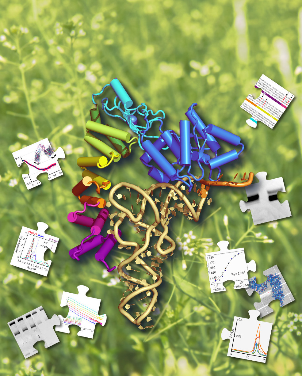
RNAs constitute a rich family of biolomecules that include for instance ribosomal, transfer, small regulatory, viral genomic RNAs or ribozymes (RNA catalysts). They are involved in processes such as the regulation of gene expression, the translation of genetic information into proteins, the maturation of other RNAs. I am particularly interested in transfer RNAs (tRNAs), which act as molecular adapters, during protein synthesis. I study their maturation, stability, and the way they interact with proteins through a multidisciplinary approach based on biochemical and biophysical methods. Indeed, beside X-ray crystallography, analyses made in solution - i.e. size exclusion chromatography (SEC), dynamic light scattering (DLS), circular dichroism (SRCD), small angle X-ray scattering (SAXS) - provide very complementary structural data to understand the properties and the multiple functions of such ribonucleoproteic systems. Among others, my work has led to the structural characterization of :
- the interaction of the aspartyl-tRNA synthetase and tRNAAsp in yeast [Sauter et al. 2000].
- the Hfq, a major chaperone of small regulatory RNAs in E. coli and other Sm-like proteins from archaea [Sauter et al. 2003].
- bacterial and mitochondrial synthetases in complex with inhibitors [Bonnefond et al. 2007, Messmer et al. 2009, Bonnefond et al. 2011, Neuenfeldt et al. 2013].
- two human mitochondrial aminoacyl-tRNA synthetases (TyrRS, AspRS) [Bonnefond et al. 2007, Neuenfeldt et al. 2013]
- the Gapevine Fanleaf virus (GFLV), an RNA virus which constitutes a major pathogen of grapevine [Schellenberger et al. 2011b, Belval et al. 2016,Orlov et al. 2020].
- the interaction of human mitochondrial AspRS and tRNAAsp [Neuenfeldt et al. 2013, Sauter et al. 2015].
- proteinaceous RNase P enzymes from Arabidopsis thaliana [Gobert et al. 2013, Pinker et al. 2017].
- strange tRNAs found in mitochondria [Messmer et al. 2009, Giegé et al. 2012, Jühling et al. 2018].
- tRNA-CCA-adding enzymes [de Wijn et al. 2018, de Wijn et al. 2019, de Wijn et al. 2020].
Biocrystallogenesis
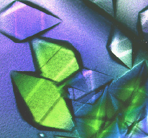
![]() Crystallography is my favorite investigation tool to study the 3D structure of biomolecules, but it requires
the production of "good" crystals. This is why
I take an interest in this field of biological
crystallogenesis, the science (the art?) of crystal growth.
Crystallography is my favorite investigation tool to study the 3D structure of biomolecules, but it requires
the production of "good" crystals. This is why
I take an interest in this field of biological
crystallogenesis, the science (the art?) of crystal growth.
Over the last twenty years, I have been involved in the development of methods that facilitate the preparation of crystals for crystallographic studies like:
- the use of phase diagrams as optimization tools [Sauter et al 1999a, Zhu et al 2002, Schellenberger et al 2011a].
- the use of additives to enhance crystal growth and quality [Sauter et al, 1999b, Bonnefond et al. 2011].
- the crystal growth in agarose gel to reach higher resolution [Sauter et al. 2002, Schellenberger et al 2011a], lower mosaicity [Lorber et al 1999a, 1999b], better cryocooling [Biertümpfel et al. 2005] or ligand soaking [Sauter et al. 2009].
- the crystallization by the counter-diffusion method [Garcia-Ruìz et al. 2001, Biertümpfel et al. 2002, Dhouib et al 2009]
- the crystallization in microfluidic chips [Sauter et al 2007, Dhouib et al 2009, Pinker et al 2013, de Wijn et al 2019]
- the preparation of microcrystals for serial crystallography [de Wijn et al 2020]
Follow me on